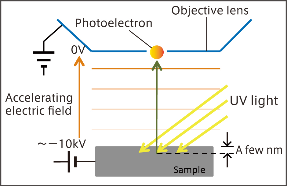
Photo Emission Electron Microscopy (PEEM)PEEM is an electron microscope that detects photoelectrons emitted from a sample by irradiating short wavelength light such as ultraviolet light.
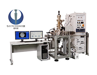
MyPEEM
PEEM is an electron microscope that detects photoelectrons emitted from a sample by irradiating short wavelength light such as ultraviolet light.
POINT
- Only general-purpose PEEM in Japan
- Visualize the status of the electron
- Electrostatic corrector is equipped
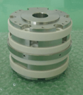
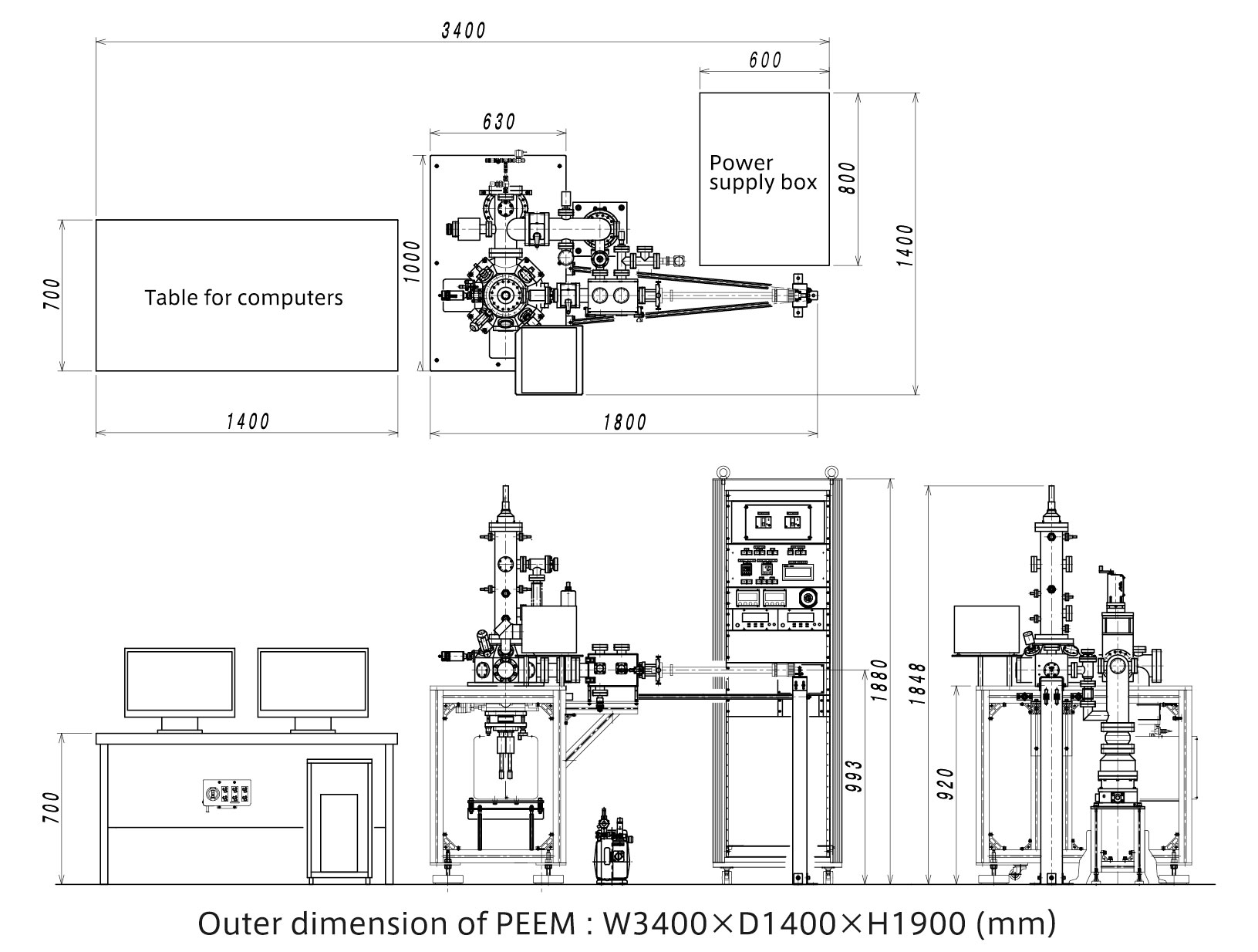
Outer dimension (Main unit)
W3400mm×D1400mm×H1900mm
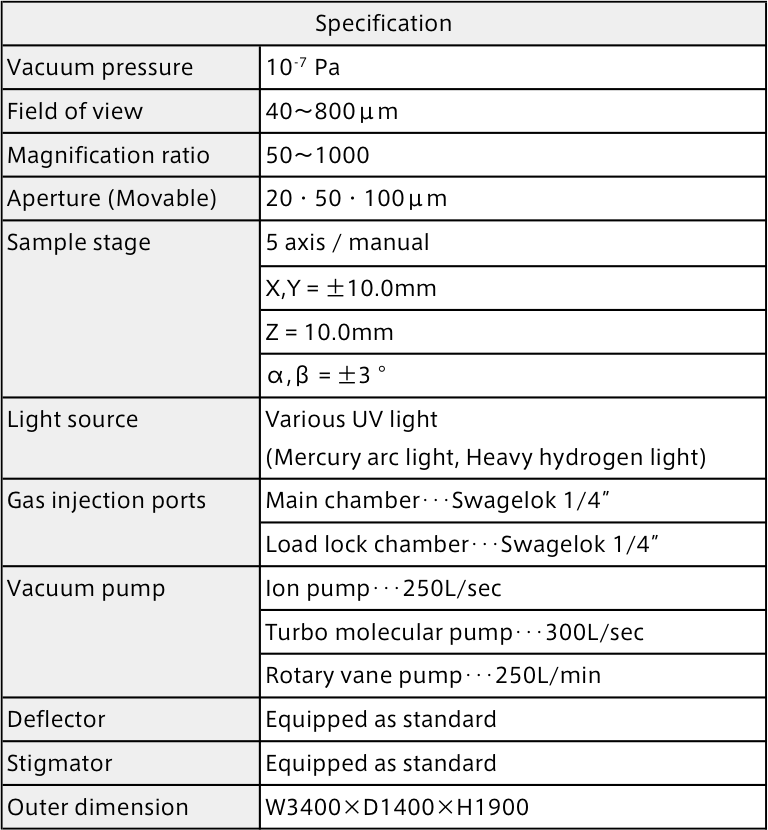
| Vacuum pressure | Field of view | Magnification ratio | Sample stage |
|---|---|---|---|
| 10-7Pa | 40~800μm | 50~1000 | 5 axis / manual |
Photo Emission Electron Microscopy (PEEM)
PEEM is an electron microscope that detects photoelectrons emitted from a sample by irradiating short wavelength light such as ultraviolet light.
Prime Minister’s Commendations
Monodzukuri Japan Award
Product/Technical development award for excellence
Patent No. 5690610
Use
Track the thin film growth processes and dynamic processes of catalytic reactions.
It can be applied to dynamic observation of surface electronic states by gas adsorption and desorption, and spin observation using circularly polarized ultraviolet light.
Features
【Feature1】Stand-alone type
This equipment integrates the PEEM itself, chamber, power supply box, and control software, so that it can be operated without any optional items.
【Feature2】Exchange of the light source
It has the configuration to irradiate the ultraviolet light as the excitation light source from outside the vacuum through the viewport so that various light sources can be easily exchanged.
【Feature3】Manual selection
It has the configuration to irradiate the ultraviolet light as the excitation light source from outside the vacuum through the viewport so that various light sources can be easily exchanged.
【Feature4】Originality
Custom made sample holder is available on demand besides the standard sample holder.
Overview
This equipment accelerates the speed of photoelectrons (secondary electrons) which are excited by irradiating the sample with ultraviolet light using the negative high voltage applied to the sample. Then, PEEM image projected on the screen using a three-stage electrostatic lens and a two-stage correction lens is recorded by CCD camera or the like.
PEEM images can be easily acquired simply by adjusting the sample height according to the field of view which is set in advance on the software.
In addition, by applying voltage to the sample, the chamber, which is the photoelectron path, can be placed at ground potential so that it has a safe and compact design.
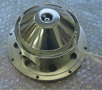
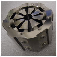
1.Electrostatic lens
This equipment consists of three-stage lens system.
Each three-stage lens system consists of 4-pole objective lens, 3-pole intermediate lens, and 5-pole projection lens. In addition, by operating the 2-pole, 1-pole, and 2-pole variable electrodes with the power operation software, the desired magnification and field of view can be obtained.
<!–
 対物レンズ |
 中間レンズ |
 投影レンズ |
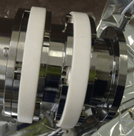
 Electrostatic corrector |
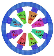 Electrostatic corrector |
3.Spatial resolution
Spatial resolution

MyPEEM Specification
MyPEEM Outer dimension
© 2024 SUGA Co., Ltd. All Rights Reserved.